December Image of the Month
Our 3 featured images in December were captured by Nicole Knight, Anderson Brock and Krista Moore!
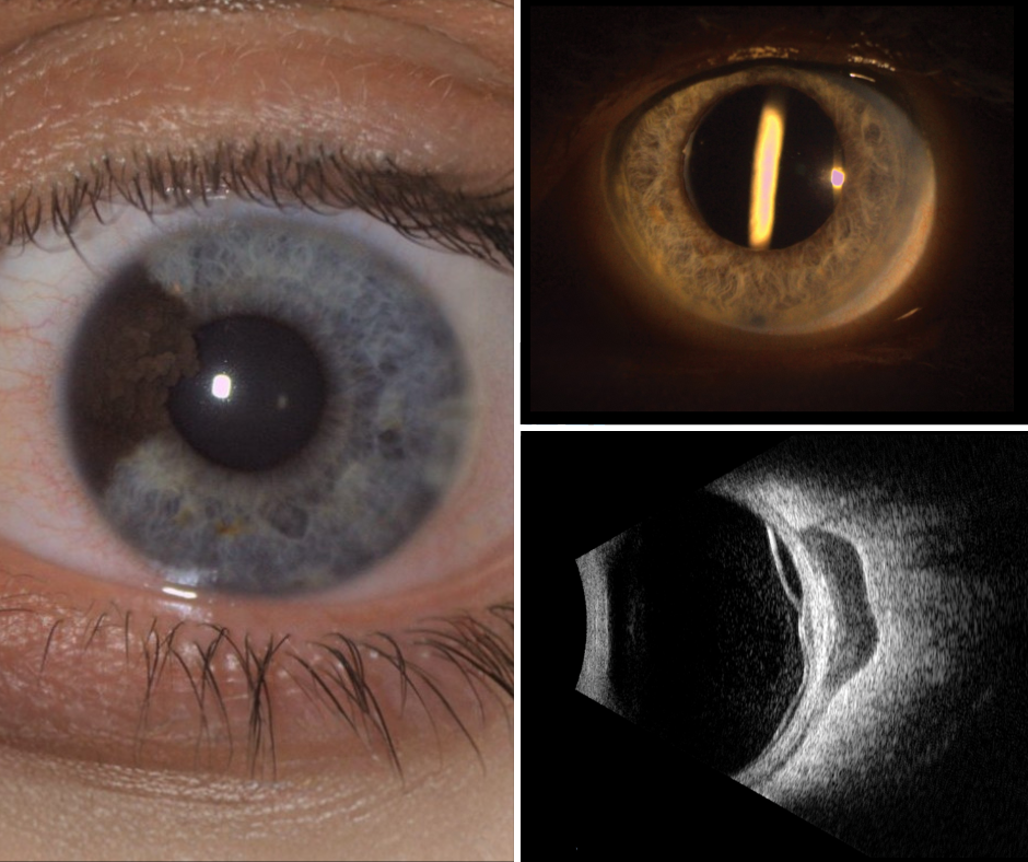
Image #1 is a slit lamp photo of an Iris Nevus captured by Nicole Knight. This image identifies that this is a raised pigmented nevus located on the iris. A nevus can be flat or raised. These are common and often referred to as a freckle or mole. It is very important to monitor a nevus with regular eye exams, since in rare cases, it can be a sign of melanoma. During your eye exam the doctor will closely monitor the nevus for changes in size, shape or color this could be a sign of melanoma. Ways to photograph and monitor your nevus during your exam include slit lamps, ultrasound and photography. Learn more about Ocular Oncology HERE
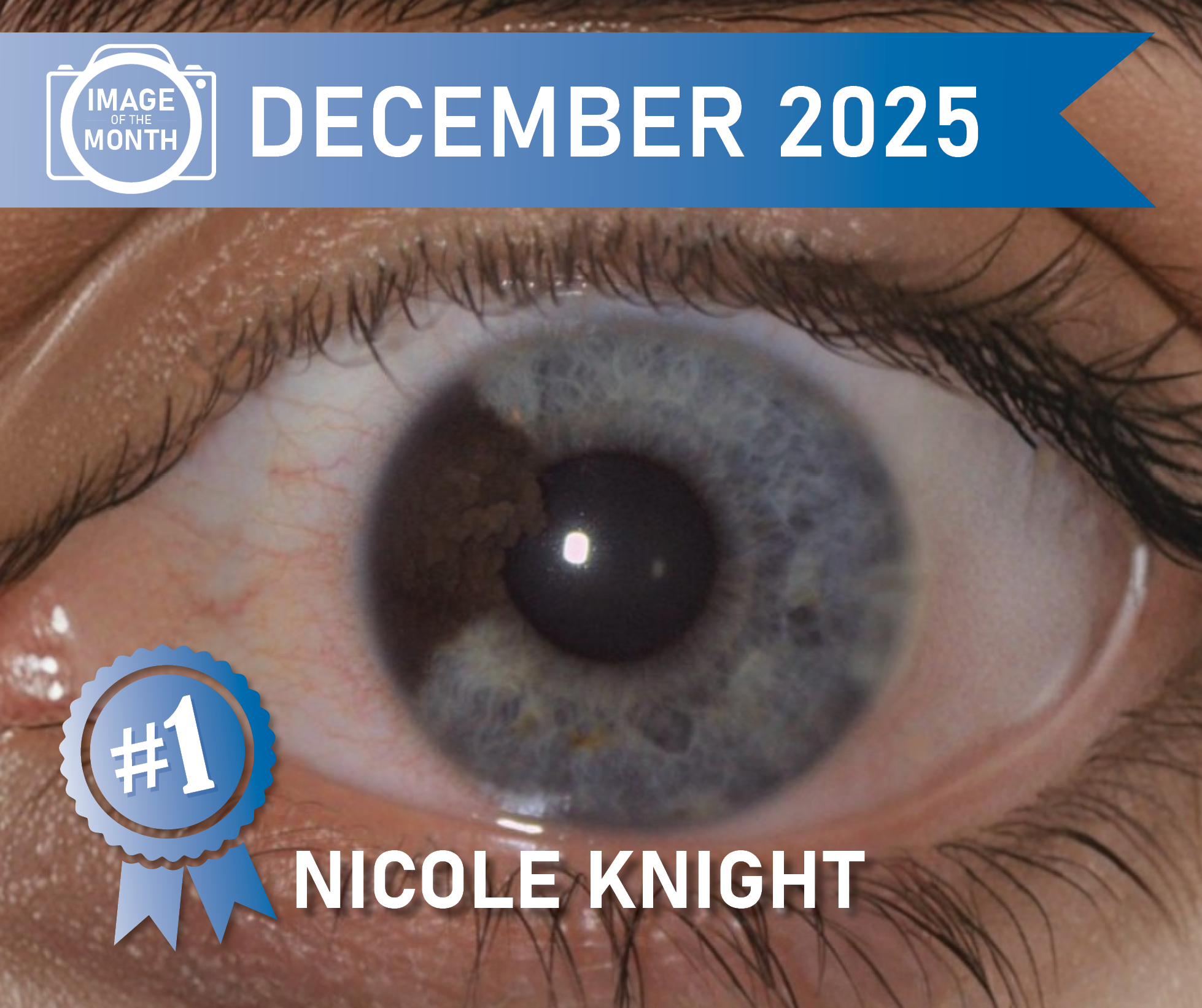
Image #2 is a photo of an Ozurdex implant that has migrated from the vitreous to against the posterior capsule, captured by Anderson Brock. This picture was taken on our slit lamp camera. This was a Uveitis patient being treated with an Ozurdex implant. Ozurdex is a small pellet that gets instilled in the eye to continuously give a dose of steroid to reduce inflammation. Read more about Uveitis HERE
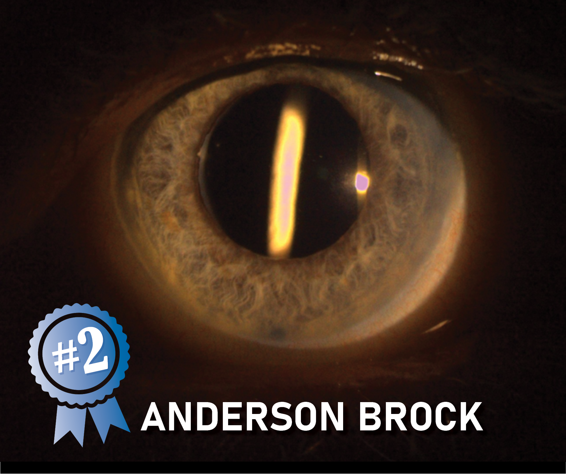
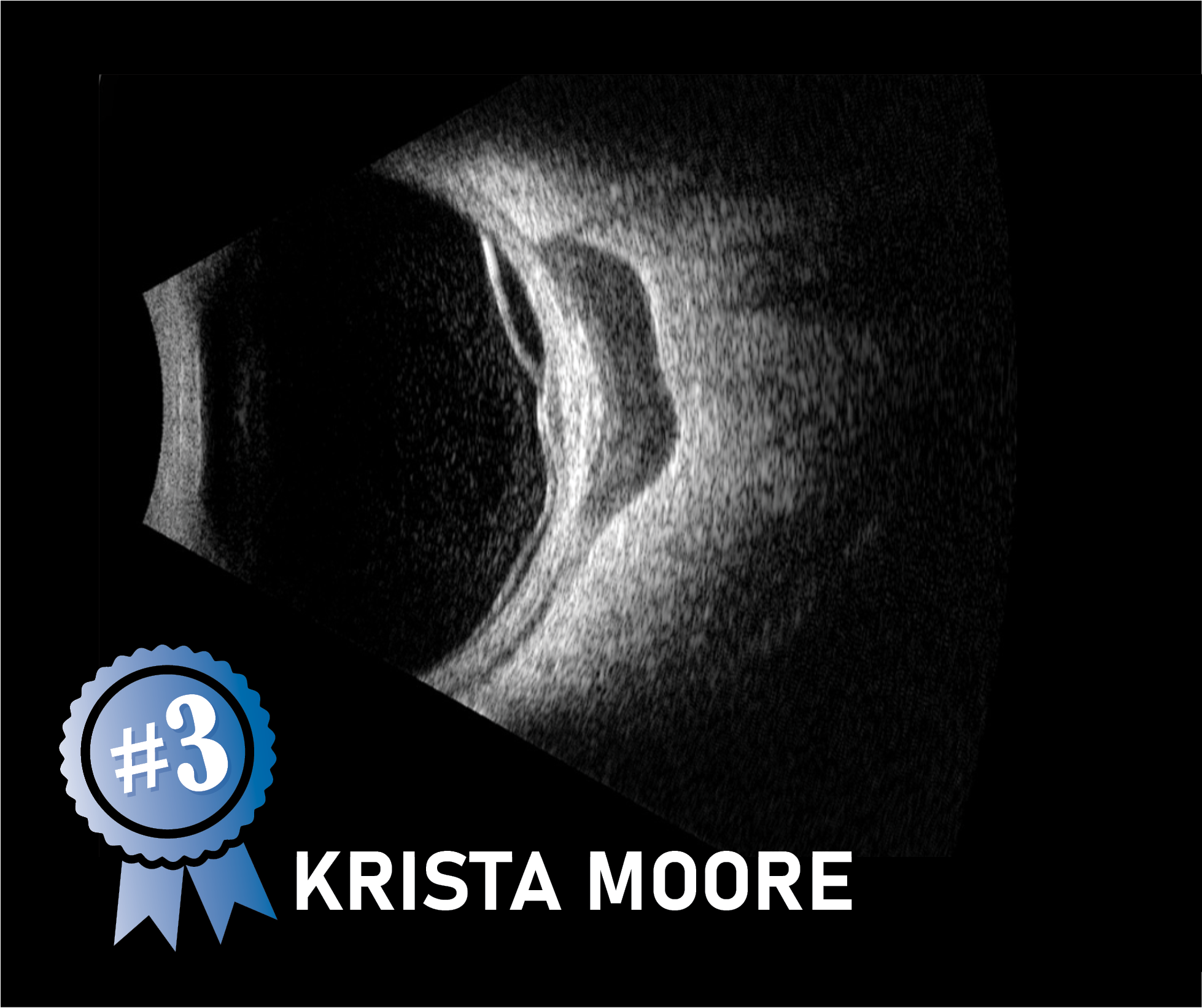
Image #3 is an ultrasound photo of Diffuse Choroidal Melanoma captured by Krista Moore. This is a type of eye cancer that grows horizontally across your choroid. This type of melanoma can be harder to detect because of its poorly defined borders, unlike the typical dome shaped tumors. Diffuse Choroidal Melanoma has a higher metastatic risk due to its larger size and difficult diagnosis. Learn more about Ocular Oncology HERE