July Image of the Month
Our 3 featured images in July were captured by Rebekah Greenberg, Krista Moore and Tiffany Del Rio!
Image #1 is a photo of a subhyaloid hemorrhage on our optical coherence tomography (OCT) machine. This hemorrhage was located in the hyaloid membrane. The posterior hyaloid membrane separates the vitreous from the retina. Symptoms of a subhyaloid hemorrhage can include sudden vision loss, floaters, and distortion.
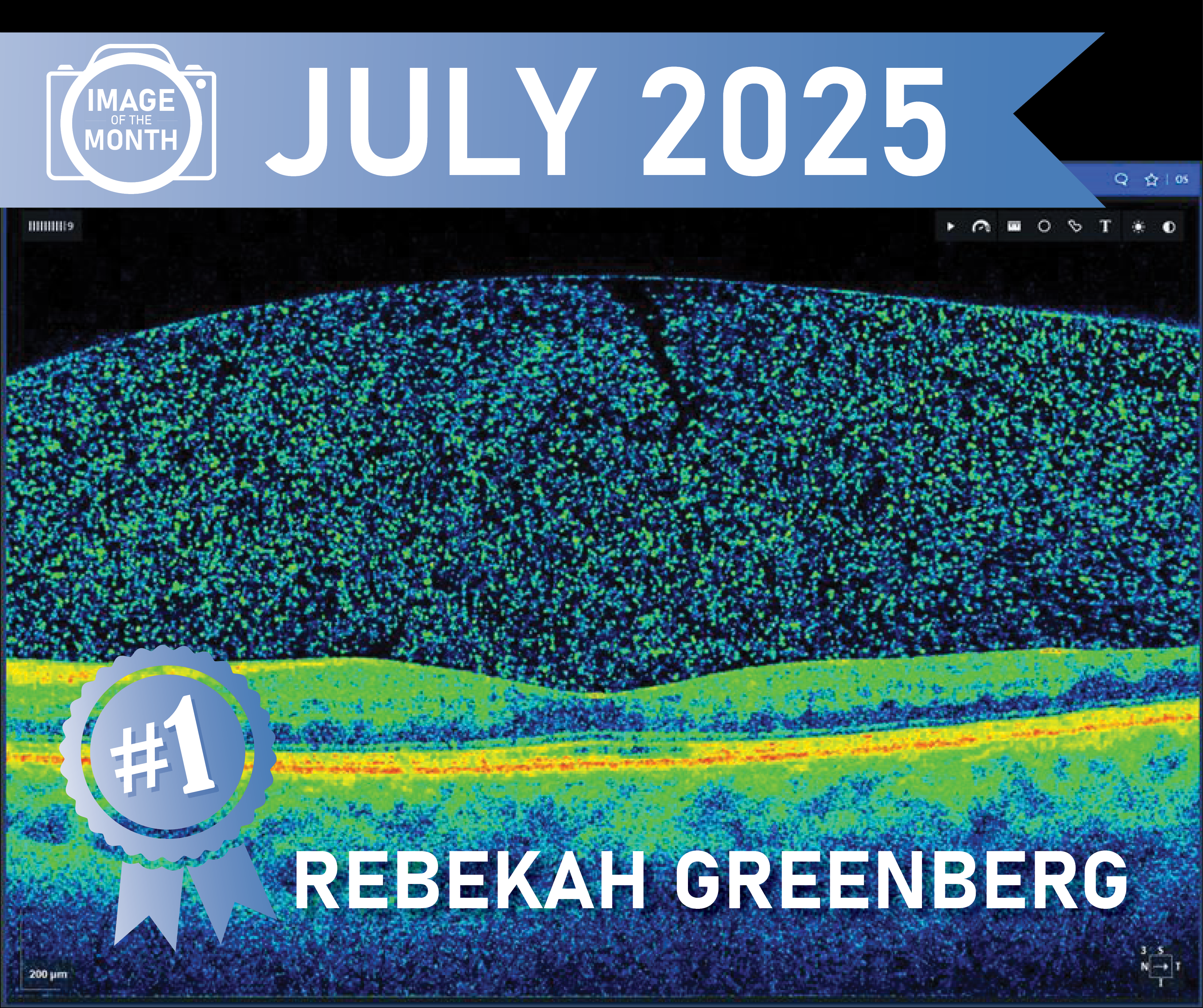
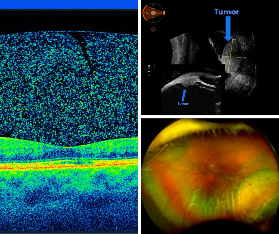
Image #2 is a photo of a Dislocated IOL and Tumor. A Dislocated IOL happens when your artificial lens that is implanted during cataract surgery moves out of its inserted position. Tumors found in the eye can be benign or malignant, further testing is crucial to identify what kind of tumor you have and for Dr Reichstein and our oncology team to put together a treatment plan. Read more about our oncology department HERE
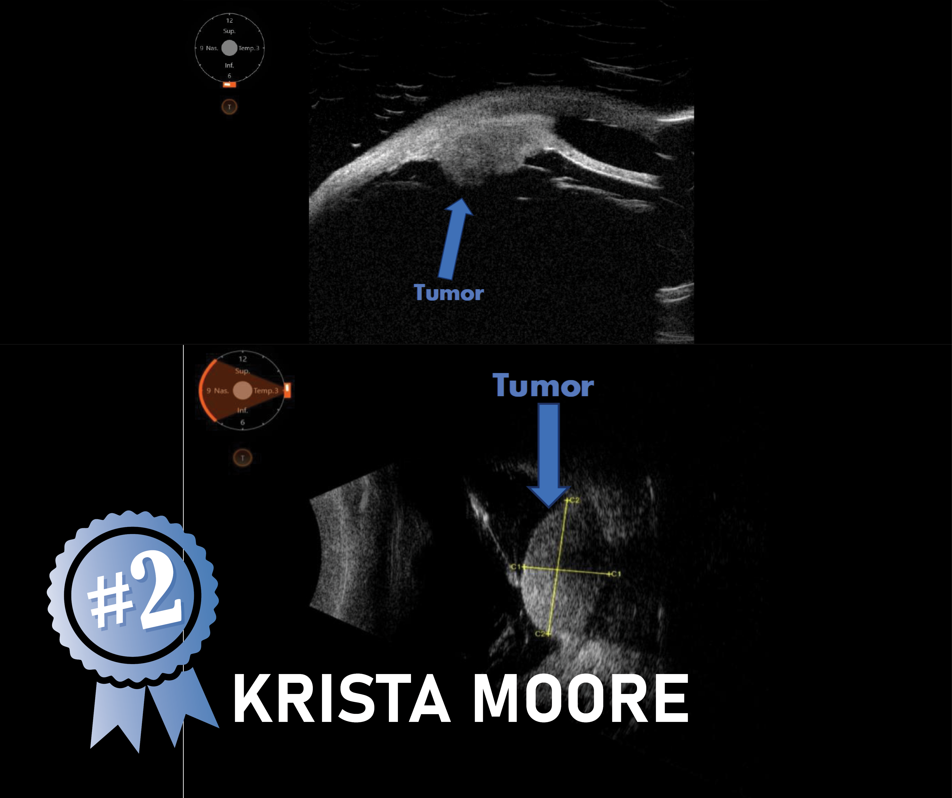
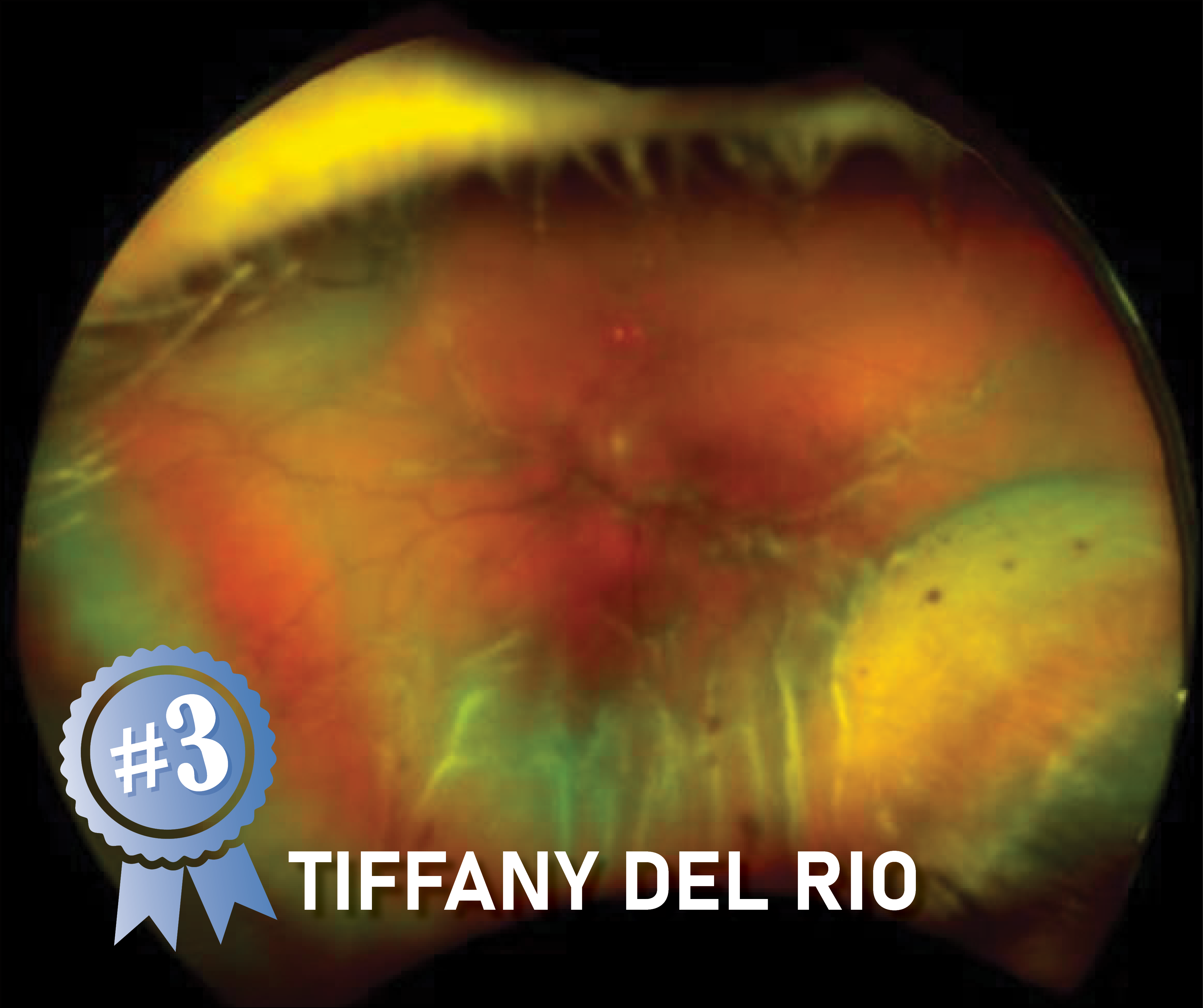
Image #3 is a photo of a Choroidal Detachment with Folds. A choroidal detachment is when you have fluid build up causing the choroid to detach from the sclera. Choroidal Folds are “wrinkles” in the retina. Read more about detachments HERE