June Image of the Month
Our 3 featured images in June were captured by Kaitlyn Anderson and Rebekah Greenberg!
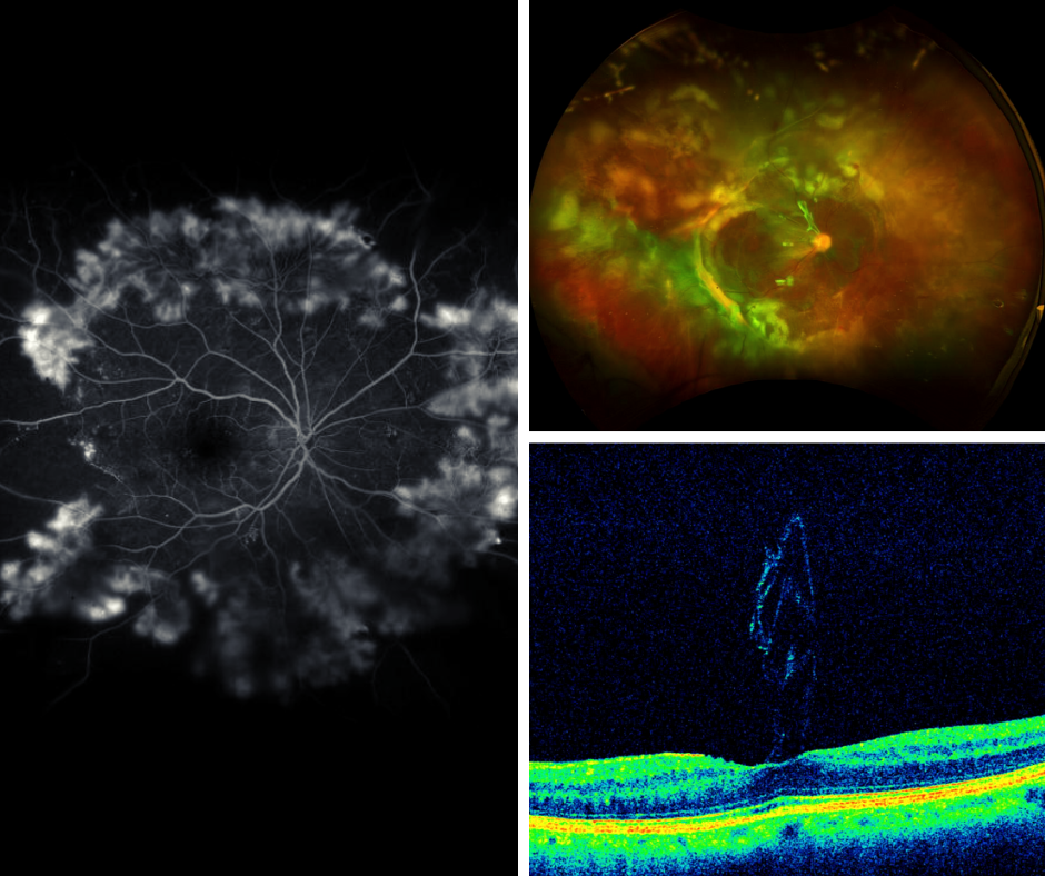
Image #1 is a photo of Proliferative Diabetic Retinopathy (PDR). PDR is a more progressed stage of Diabetic Retinopathy and is characterized by the growth of new, abnormal blood vessels (neovascularization) on the retina. The abnormal blood vessels often hemorrhage (bleed) into the eye and can cause scar tissue on the retina and other complications within the eye. Learn More

Image #2 is a photo of a Tractional Retinal Detachment (TRD). Tractional Detachments are caused by scar tissue that contracts and causes the retina to pull away from the important retinal pigment layer in the back of the eye. This type of detachment is most common in patients with diabetes. Learn More

Image #3 shows an unusual presentation of an Epiretinal Membrane (ERM)—note how the membrane appears to "stand up" rather than lie flat against the retina. ERMs, or macular puckers, typically form as a thin, scar-like tissue on the surface of the macula, causing distortion or blurring of central vision. While most develop spontaneously, some are linked to underlying eye conditions. This scan highlights just how varied ERM presentations can be. Learn More