November Image of the Month
Our 3 featured images in November were captured by Nicole Knight, Tiffany Del Rio and Jerome Cole!
Image #1 is an ultrasound photo of an Exudative Retinal Detachment. This B scan was captured by Nicole Knight. A B scan is an ultrasound image that is created by a 2D cross-sectional picture of an internal body structure. This scan uses sound waves to detect problems such as a retinal detachment. Exudative retinal detachments form when fluid is leaked out of blood vessels and accumulates under the retina. This type of retinal detachment is much less common and can occur in eyes with abnormal inflammation or excessive leakage from abnormal blood vessels.
Read more about Retinal Detachments HERE

Image #2 is a photo of an Inactive Stable Serpiginous Lesions captured by Tiffany Del Rio. These findings can be associated with Serpiginous Chorioretinitis and Uveitis. She captured this image by capturing an auto fluorescence and color fundus photo. Uveitis is a condition characterized by inflammation in the eye. Symptoms of uveitis can vary based on the location of predominant inflammation. About 40% of uveitis cases may not have an underlying case (idiopathic). Read more about Uveitis HERE
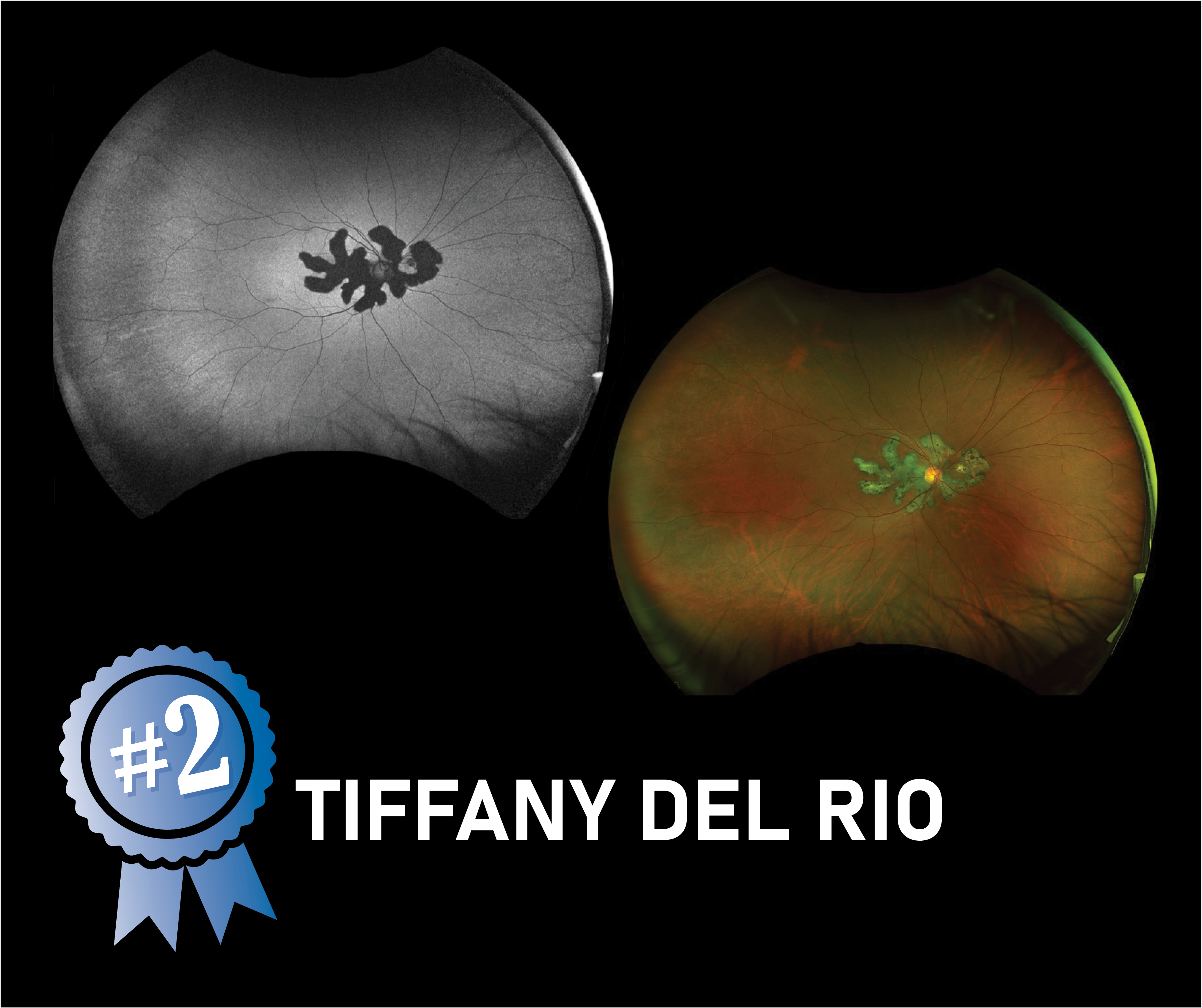
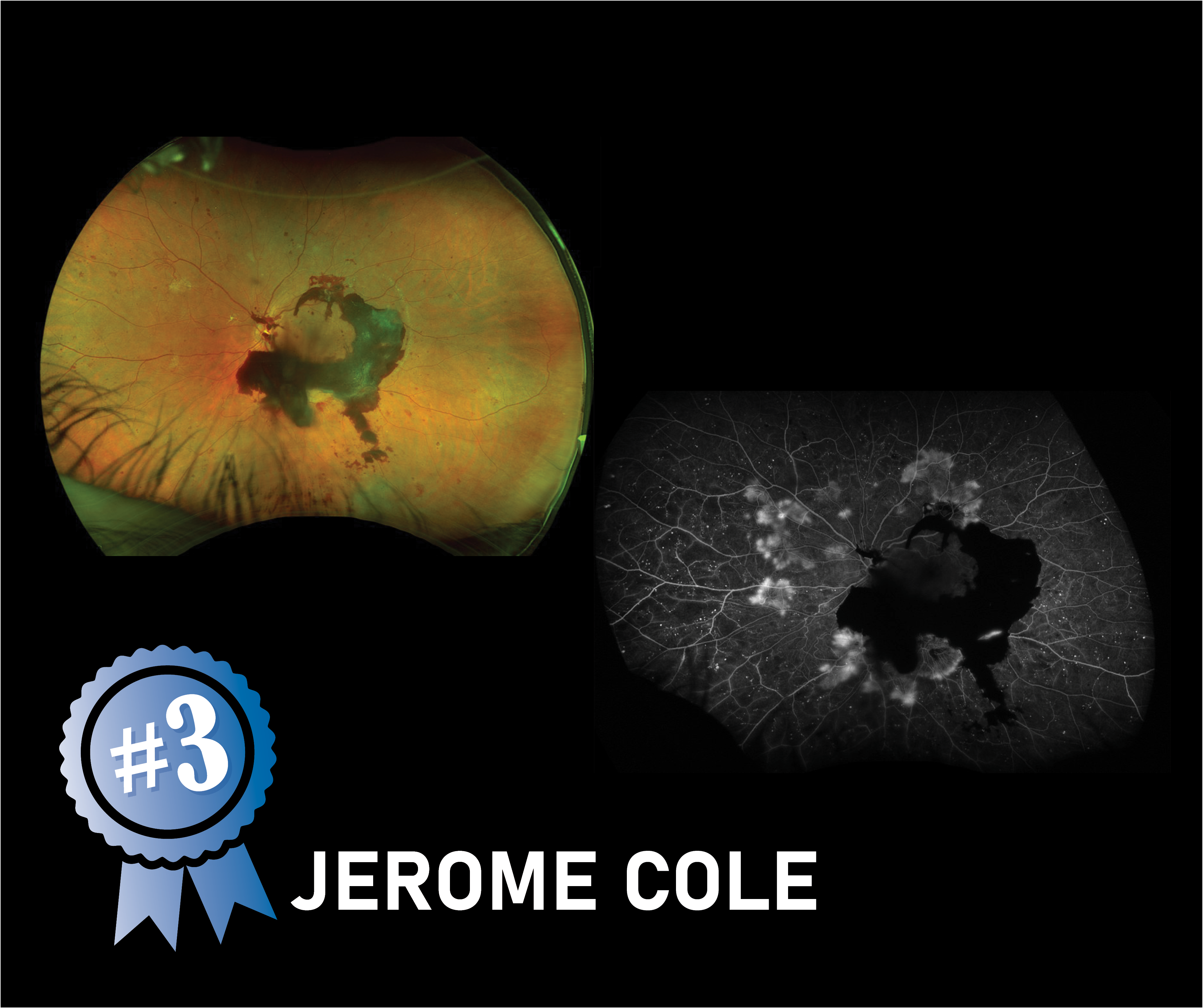
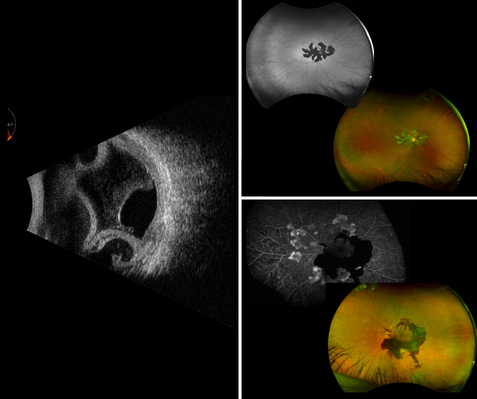
Image #3 is a Vitreous Hemorrhage and Neovascularization Elsewhere (NVE) captured by Jerome Cole. This can be commonly seen in patients diagnosed with Severe Diabetic Retinopathy. This photo was taken using our OPTOS Camera. Diabetic Retinopathy (DR) is one of the most common causes of vision loss. High blood sugar levels cause damage to the small blood vessels in the retina. Read more about Diabetic Retinopathy HERE