September 2026 Image of the Month
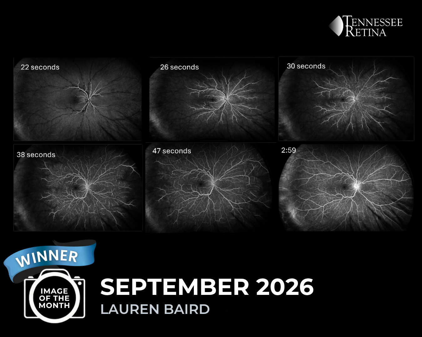
Our 3 featured images in September were captured by Lauren Baird , Carla Sensabaugh and Anita Bingaman!
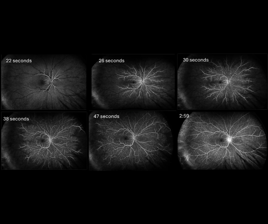
Image #1 is a fluorescein angiogram photo of an impending central retinal vein occlusion taken on our OPTOS camera by Lauren Baird. Findings include dilated tortuous vessels, and patchy areas of ischemia (scattered, localized areas where tissues and organs are not getting enough oxygen rich blood). CRVO (Central Retinal Vein Occlusion) is typically a clinical diagnosis that is, one based on medical signs and patient reported symptoms. When a retina specialist looks into the eye, there is a characteristic pattern of retinal hemorrhages (bleeding) and a diagnosis is made. Read more about CRVO’s HERE
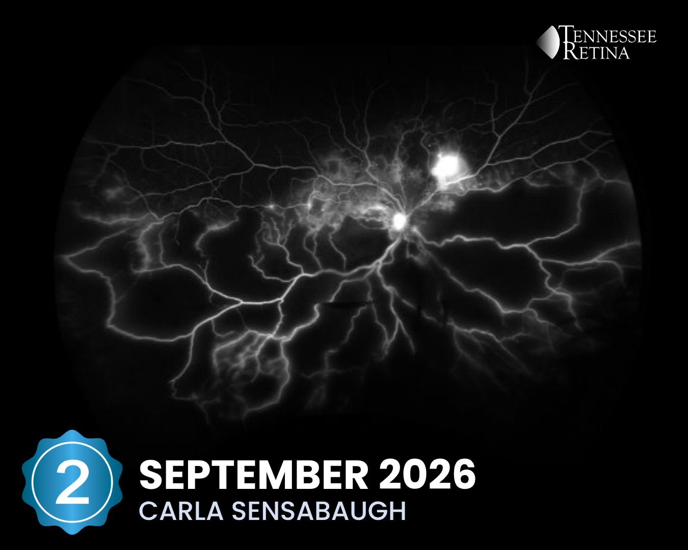
Image #2 shows capillary nonperfusion in a Fluorescein Angiogram that is related to Diabetes. Carla Sensabaugh took this photo on our Optos Camera. Capillary nonperfusion is a complication from diabetes that results in the blockage or closure of small blood vessels in the retina, that will cause part of the eye to be deprived from oxygen and blood flow. Learn more about how diabetes can affect our eyes HERE
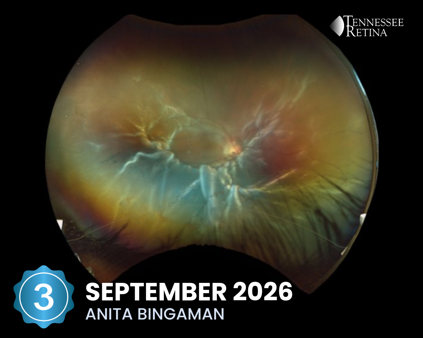
Image #3 is a color fundus photo of a total Retinal Detachment taken by Anita Bingaman. The retina is the inner lining of the eye; it is the thin, light-sensitive tissue that generates vision. Tears can form in the retina, creating a risk of retinal detachment and severe loss of vision. If a retinal tear is diagnosed promptly before it progresses to retinal detachment, the prognosis is extremely good. Learn more HERE