October 2026 Image of the Month
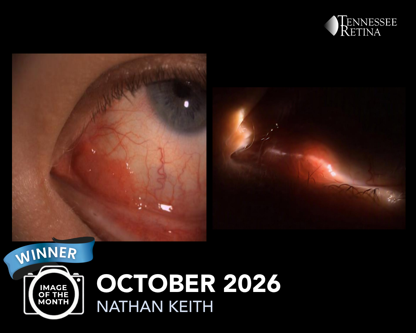
Our 2 featured images in October were captured by Nathan Keith and Anita Bingaman!
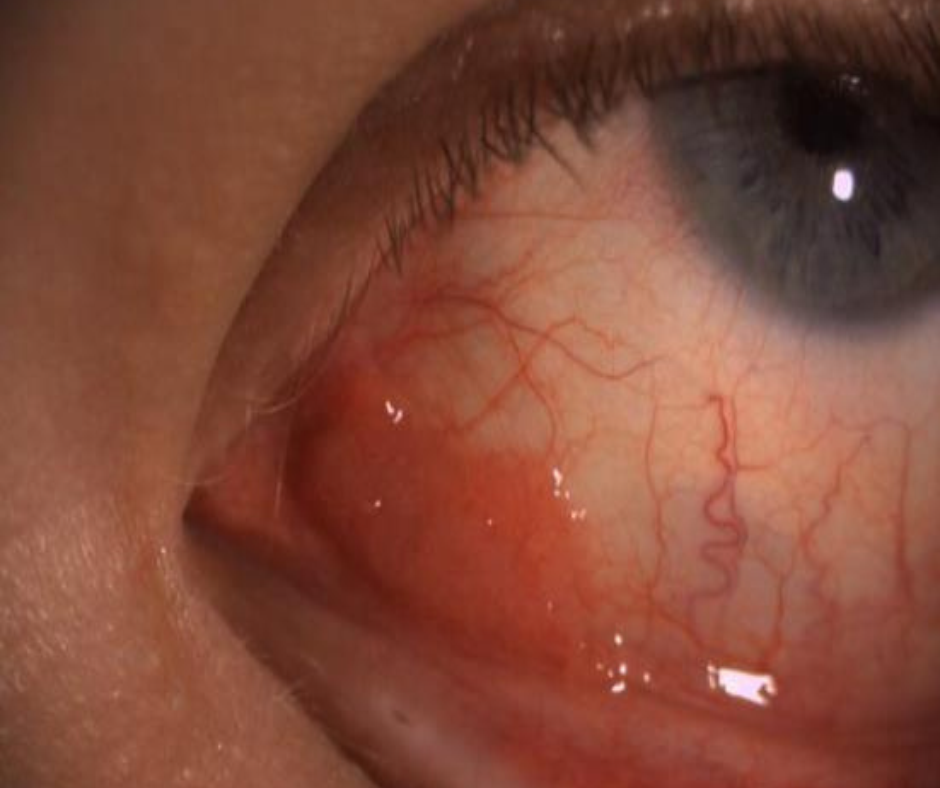
Image #1 is picture of Squamous Cell Carcinoma taken by Nathan on our Slit Lamp Camera. A visible finding included a Gelatinous (thick, viscous, and jelly like consistency) fibrotic pink lesion. This type of growth can develop in many different parts of the eye, including the eyelid, conjunctiva, and lacrimal sac. Depending on size and location, Squamous Cell Carcinoma can be treated with surgery, cryotherapy or radiation therapy. Read more about our Ocular Oncology department HERE
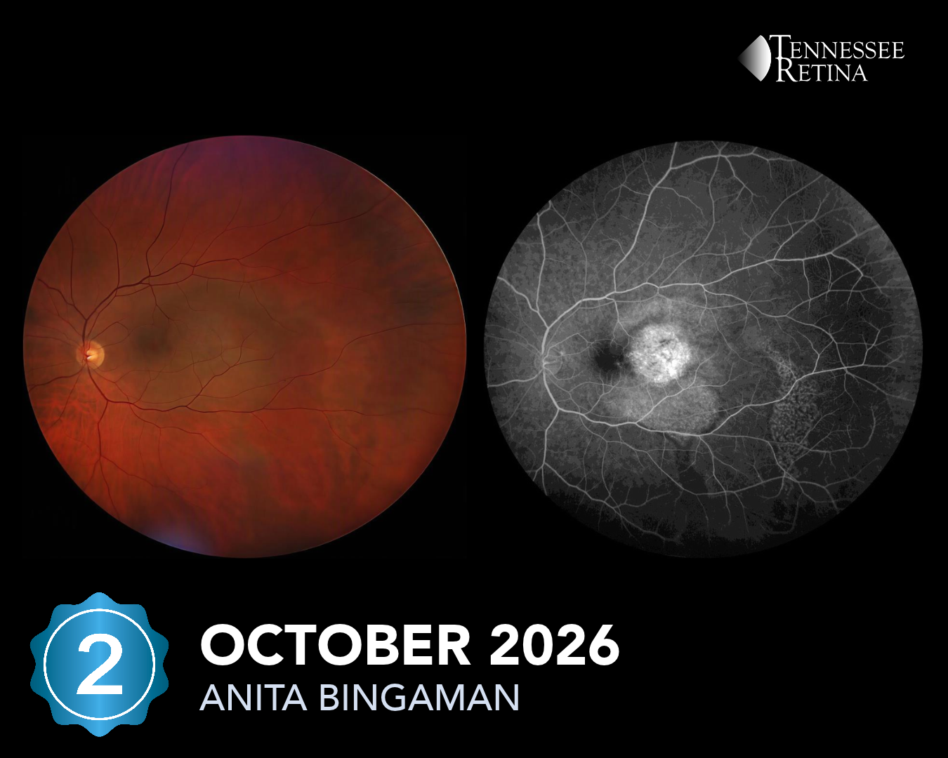
Image #2 captured by Anita, is a photo of a Choroidal Hemangioma. This type of Hemangioma develops inside the eye in the layer that holds blood vessels, located behind the retina. These can sometimes cause blurry vision, fluid buildup, or retinal changes. Treatment of a Hemangioma depends on whether or not it is affecting your vision.